An umbilical hernia is a condition where a bulge appears at the belly button. It happens when a small part of the intestine or fatty tissue pushes through a weak spot in the abdominal wall muscles. In most cases, these hernias are harmless and don’t cause any symptoms.
Most umbilical hernias resolve spontaneously by the age of 3 years. For this reason, the standard approach for asymptomatic hernias is “watchful waiting.” There is no evidence that taping or binding the hernia helps, and it can even cause skin irritation.
Surgical repair is typically recommended for hernias that:
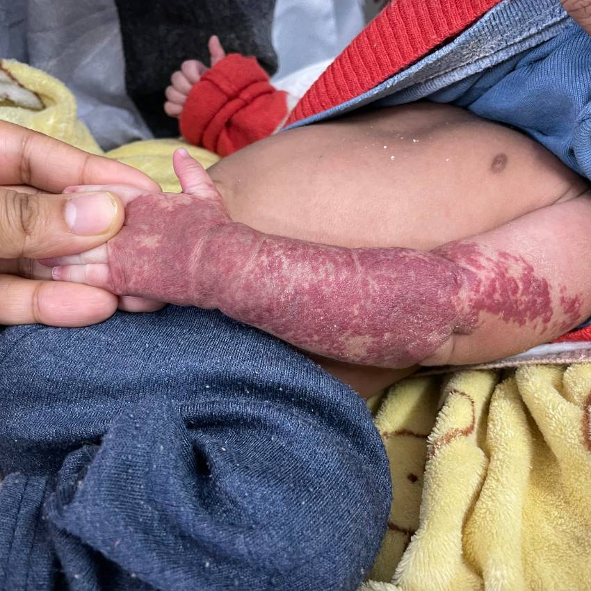
Vascular anomalies are birthmarks or growths caused by abnormal blood vessels. The most common type is an infantile hemangioma, which often appears as red, raised spots (like a strawberry) that grow rapidly in the first year of life before shrinking on its own. They usually don’t require treatment unless they interfere with a child’s vision, breathing, or cause bleeding.
Other types of vascular malformations are present at birth and grow proportionally with the child and may look like large, disfiguring bluish masses. They don’t go away on their own. Management options include observation, compression therapy, sclerotherapy, laser therapy, or surgery. The goal of management is to relieve symptoms and prevent complications, improving the child’s quality of life.
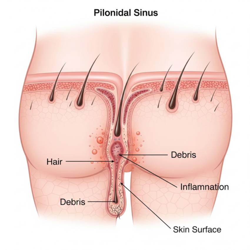
A pilonidal sinus is a small pit or tunnel that develops in the skin at the top of the buttocks crease caused by a combination of factors, including ingrown hair, friction, and pressure on the area. It can become infected to cause a painful abscess (a collection of pus).
Management includes drainage of the abscess to remove the pus and relieve the pain. After the infection has cleared, the pilonidal sinus itself requires a surgical procedure to remove the entire tract and cavity to prevent it from becoming infected again and again.
Polydactyly and syndactyly are two of the most common congenital hand and foot anomalies in children. Polydactyly is the presence of one or more extra fingers or toes whereas Syndactyly is the fusion of two or more fingers or toes.
The management for both conditions is typically surgical. For polydactyly, removal of the extra digit is done with timing of surgery depending on the location and size of the extra digit. For syndactyly, surgery is performed to separate the fused digits, often requiring skin grafts. The timing of surgery is carefully considered to allow for normal hand function as the child grows.