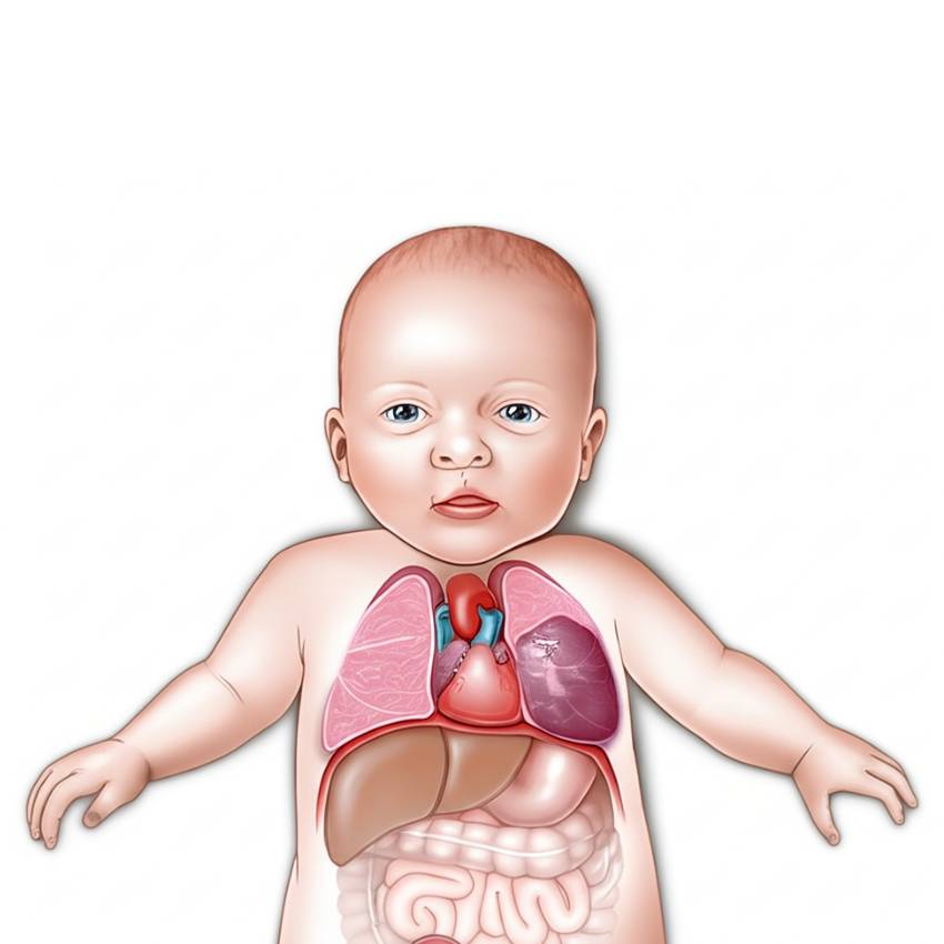
Cystic lung lesions are a group of rare, non-cancerous malformations of the respiratory tract that occur during foetal development. They are essentially abnormal areas of lung tissue that do not function correctly.
While many cases are detected on prenatal ultrasound, some lesions may also present after birth with breathing difficulties or recurrent, pneumonia.
Treatment, for most cases, involves surgical removal of the abnormal tissue (lobectomy), either via open or thoracoscopic surgery.
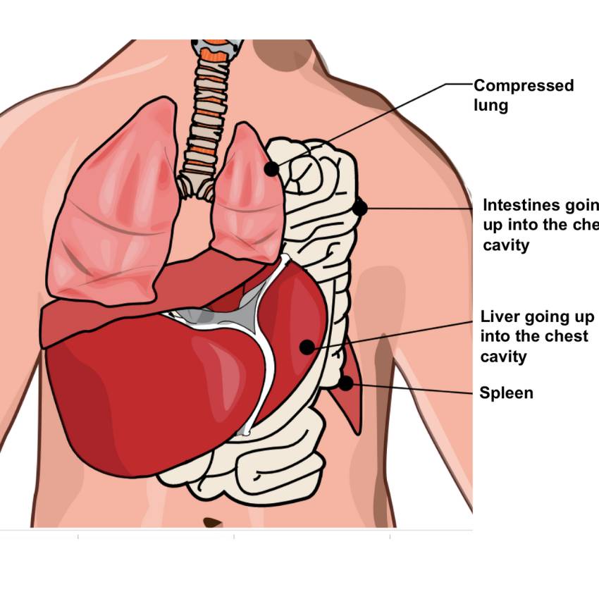
CDH is a birth defect where a hole in the diaphragm, the muscle separating the chest and abdomen, allows abdominal organs (like the stomach, liver, and intestines) to move into the chest cavity. This is a life-threatening condition as it prevents the lungs from developing properly.
CDH is often diagnosed on prenatal ultrasound. After birth, infants typically present with severe breathing distress.
The immediate treatment involves stabilizing the baby’s breathing in the newborn ICU. This is followed by surgical repair of the diaphragm, which involves moving the abdominal organs back into the abdomen and closing the hole.
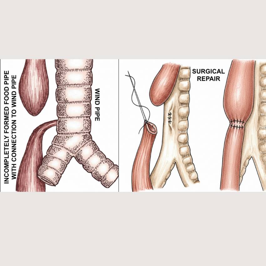
Oesophageal atresia and tracheo-oesophageal fistula (OA/TOF) are birth defects affecting the oesophagus (food pipe) and trachea (windpipe) where the food pipe is incomplete and there is an abnormal connection between the food and the windpipe. This is diagnosed when babies have difficulty in feeding after birth or have frothy, white bubbles in the mouth.
This is a medical emergency that requires immediate attention in the newborn ICU. Management involves stopping all oral feeding and using a suction tube to keep the baby’s airway clear. Surgical repair is performed soon after birth to connect the two ends of the oesophagus and close the abnormal connection.
With modern medical care and surgical techniques, most babies have successful surgeries and go on to live normal, healthy lives.
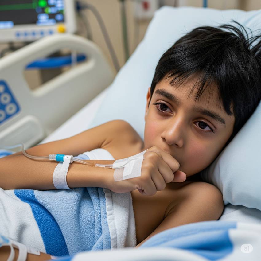
Empyema is a collection of pus in the pleural space (around the lungs). Children can present with persistent fever, cough, and shortness of breath or it might be secondary to pneumonia or tuberculosis.
Management involves a multi-pronged approach. Initial steps include appropriate antibiotic therapy and sometimes, chest tube insertion to drain the thick pus. In some cases, a more aggressive approach like video-assisted thoracoscopic surgery (VATS) may be required to break up loculations and fully drain the pus.