Lymphatic malformations (LM) are benign growths of the lymphatic system, which can occur anywhere in the body, but are commonly found in the neck and head region of newborn babies. They are thought to arise from an error in the development of the lymphatic vessels, leading to the formation of fluid-filled sacs or cysts.
Larger swellings can compress vital structures, such as the trachea (windpipe) and oesophagus (foodpipe), leading to breathing and feeding difficulties. They can also cause cosmetic deformity.
Treatment options range from monitoring and waiting for spontaneous regression to sclerotherapy (injecting a medicine into the cysts to shrink them) to surgical excision in rare situations.
Cystic lung lesions are a group of rare, non-cancerous malformations of the respiratory tract that occur during foetal development. They are essentially abnormal areas of lung tissue that do not function correctly.
While many cases are detected on prenatal ultrasound, some lesions may also present after birth with breathing difficulties or recurrent pneumonia.
Treatment, for most cases, involves surgical removal of the abnormal tissue (lobectomy), either via open or thoracoscopic surgery.
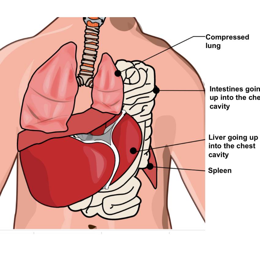
CDH is a birth defect where a hole in the diaphragm, the muscle separating the chest and abdomen, allows abdominal organs (like the stomach, liver, and intestines) to move into the chest cavity. This is a life-threatening condition as it prevents the lungs from developing properly.
CDH is often diagnosed on a prenatal ultrasound. After birth, infants typically present with severe breathing distress.
The immediate treatment involves stabilising the baby’s breathing in the newborn ICU. This is followed by surgical repair of the diaphragm, which involves moving the abdominal organs back into the abdomen and closing the hole.
Abdominal wall defects are birth problems where a baby’s intestines or other organs protrude through a hole on the tummy.
These are usually detected during prenatal ultrasounds. At birth, they are immediately visible.
Treatment involves surgical repair, often soon after birth. For large defects, the organs may be placed in a “silo,” a protective plastic bag, and gradually pushed back into the abdomen over several days before the final surgical closure.
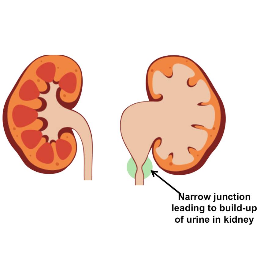
Hydronephrosis is a condition where one or both of a baby’s kidneys become swollen due to a buildup of urine. This happens when there is a blockage in the urinary tract (often known as PUJO) or when urine flows backward from the bladder into the kidney (vesicoureteral reflux).
Often, hydronephrosis is diagnosed during a prenatal ultrasound, or it may be picked up later in life if associated with abdominal swelling or urinary tract infection.
Treatment depends on the severity and underlying cause. Mild cases often resolve on their own and are managed with close monitoring via ultrasound. For more severe cases, surgical intervention may be necessary to correct the blockage or reflux to prevent long-term kidney damage.
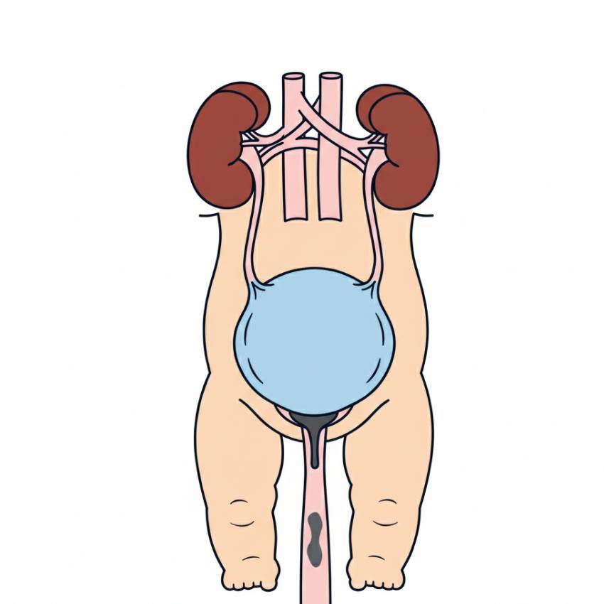
Posterior urethral valves (PUV) are a rare congenital condition affecting only baby boys where an abnormal flap of tissue in the urethra (urine tube) blocks the flow of urine out of the bladder. The blockage causes urine to back up into the bladder and kidneys, leading to significant damage.
PUV is often diagnosed on a prenatal ultrasound. At birth, boys with PUV can present with a weak urinary stream, straining to urinate or urinary tract infection.
Treatment is always surgical where a tiny scope (camera) is inserted into the urethra to surgically cut the valve tissue. Timely diagnosis and treatment are crucial to prevent permanent kidney and bladder damage.
Finding out your baby has a structural anomaly in the kidney or genital organs can be concerning, but it’s important to know that many of these conditions are manageable.
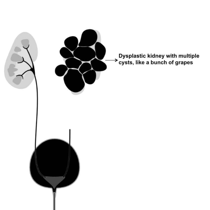
One such condition is a multicystic dysplastic kidney (MCDK), where a kidney develops as a cluster of cysts and doesn’t function. In most cases, the other kidney is healthy and compensates completely. No treatment is usually needed, but your baby will be monitored after birth to confirm the diagnosis and ensure the other kidney is healthy.
Another anomaly is a duplex collecting system, where a kidney has two ureters instead of one. This can sometimes cause urinary reflux or obstruction, but often it causes no problems at all. If treatment is needed, it might involve medication or, in rare cases, a surgical procedure to correct the issue.
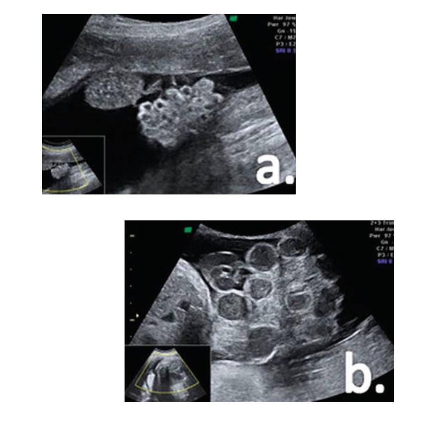
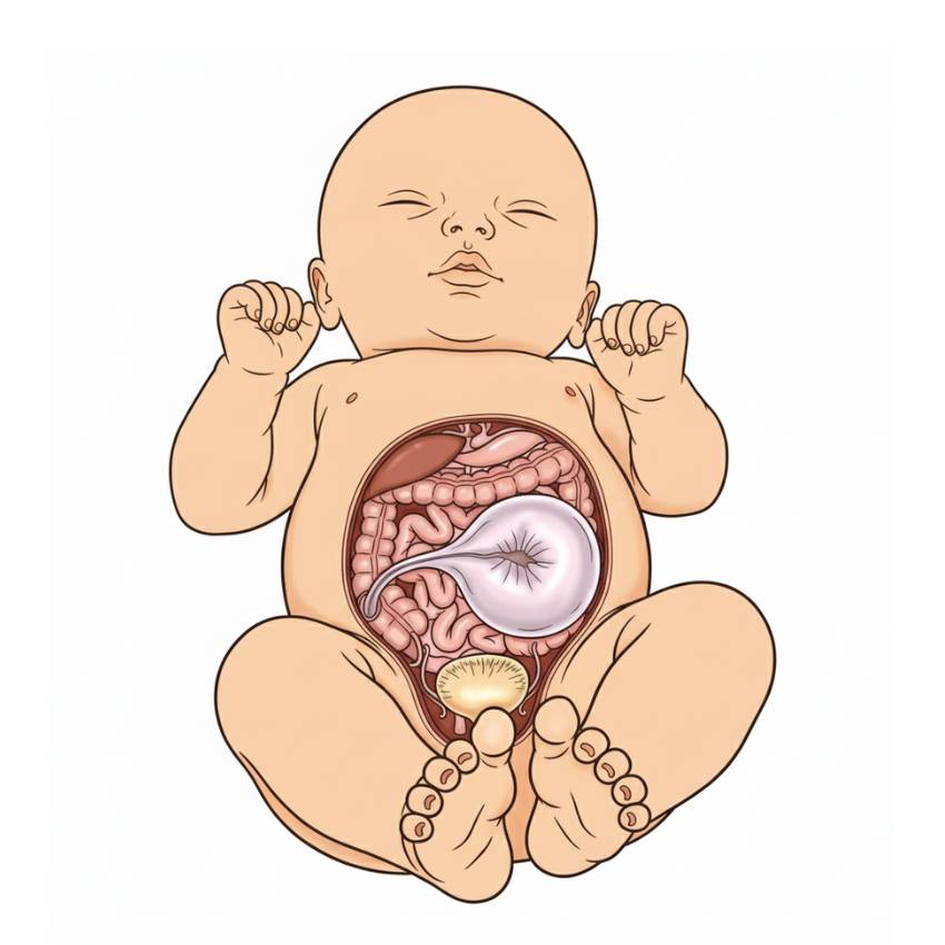
Abdominal cysts appear as fluid-filled sacs within the foetal abdomen. These can originate from various organs, including the ovaries, kidneys, mesentery, or gastrointestinal tract.
Management is largely dependent on the size and characteristics of the cyst. The majority of cysts are benign and resolve spontaneously, often before or shortly after birth. After the baby is born, small, asymptomatic cysts are usually monitored with serial ultrasounds. For larger cysts, there is a higher risk of complications such as torsion (twisting of the cyst), which can cause pain and damage to the organ. In these cases, surgical removal may be required.
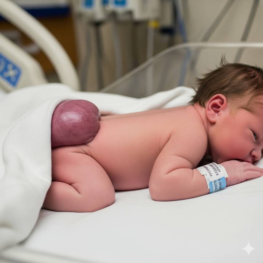
A sacrococcygeal teratoma (SCT) is a rare type of tumour that develops at the base of a baby’s tailbone. These tumours are made of different types of tissue, like hair, muscle, or bone, and can be either benign (non-cancerous) or, less commonly, malignant (cancerous).
SCTs are typically discovered during a prenatal ultrasound as a mass at the bottom of the spine. Large tumours can be a concern because they can steal blood flow from the baby, leading to heart problems or hydrops (excess fluid buildup).
Management depends on the size and type of the tumour. Small, benign tumours are usually monitored. Larger tumours or those that show signs of problems often require delivery in a specialised hospital. After birth, the tumour has to be surgically removed.